Series The new image
Stromal lymphocytes - coloured
2025
C-print, 82× 50 cm
The new image
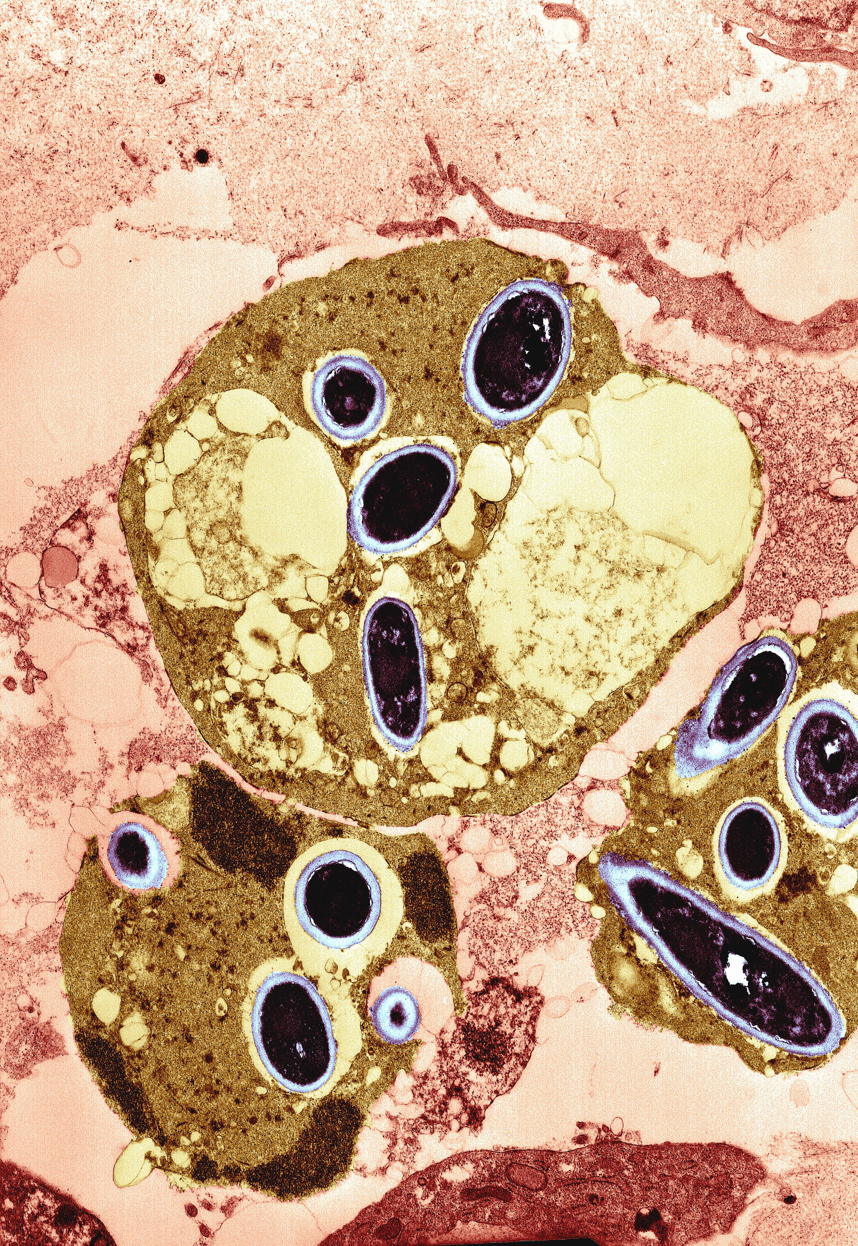
Colour-enhanced, transmission electron micrograph of a mass of small lymphocytes in the conjunctiva of a patient with an autoimmune ulceration of the cornea.
The cells have large nuclei and numerous mitochondria. A centriole can be seen towards the centre left of the centre top cell. Collagen fibrils (green) are visible in the stroma between the cells. courtesy Wellcome Collection